

| 中文名称 | 甲磺酸萘非那韦 |
| 英文名称 | NELFINAVIR |
| CAS号 | 159989-64-7 |
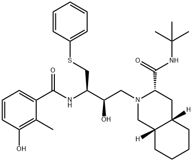
| 分子式 | C32H45N3O4S |
| 分子量 | 567.78 |
| EINECS号 | 1533716-785-6 |
| 熔点 | 185-186 °C |
| 比旋光度 | D -119.23° (c = 0.26 in methanol) |
| 沸点 | 786.8±60.0 °C(Predicted) |
| 密度 | 1.22±0.1 g/cm3(Predicted) |
| 溶解度 | 乙醇中≥20.45mg/mL |
| 形态 | 粉末 |
| 酸度系数(pKa) | pKa1 6.0; pKa2 11.06(at 25℃) |
| 颜色 | 白色至米白色 |
| 水溶解性 | 7g/L(temperature not stated) |
| 毒性 | rat,LD,oral,> 5gm/kg (5000mg/kg),Toxicologist. Vol. 42, Pg. 55, 1998. |
|
HIV-1
|
Nelfinavir (AG1341) (1-10 μM; 48 hours) inhibits the proliferation of multiple myeloma cells.
Nelfinavir inhibits 26S chymotrypsin-like proteasome activity, impairs proliferation and triggers apoptosis of the myeloma cell lines and fresh plasma cells.
Nelfinavir (1-10 μM; 17 hours) induces apoptosis of multiple myeloma cell lines.
Nelfinavir (5 μM; 0-24 hours) decreases the phosphorylation of AKT.
Nelfinavir activates the cleavage of caspase-3, decreases the phosphorylation of AKT, STAT-3, ERK1/2, and activates the pro-apoptotic pathway of the unfolded protein response system.
Cell Proliferation Assay
| Cell Line: | RPMI, LP1, U266, OPM2 and MM1S cells |
| Concentration: | 1, 2, 5, 10 μM |
| Incubation Time: | 48 hours |
| Result: | Inhibited the proliferation of RPMI, LP1, U266, OPM2 and MM1S cell lines in a dose-dependent manner with an IC 50 of 1-5 μM. |
Apoptosis Analysis
| Cell Line: | LP1 and U266 cells |
| Concentration: | 1-10 μM |
| Incubation Time: | 17 hours |
| Result: | Induced a dose-dependent increase in the percentage of annexin V + /propidium iodide + cells. |
Western Blot Analysis
| Cell Line: | U266 cells |
| Concentration: | 5 μM |
| Incubation Time: | 0-24 hours |
| Result: | The level of AKT phosphorylation in U266 cells decreased. |
Nelfinavir (AG1341) (75 mg/kg; i.p.; 5 days a week for 21 days) decreases multiple myeloma cell growth in NOD/SCID mice.
| Animal Model: | NOD/SCID mice (bearing U266-luc cells) |
| Dosage: | 75 mg/kg |
| Administration: | I.p.; 5 days a week for 21 days |
| Result: | Decreased MM cell growth in NOD/SCID mice. |
-
CAS号:155213-67-5
利托那韦 -
CAS号:198904-31-3
阿扎那韦 -
CAS号:191114-48-4
泰利霉素 -
CAS号:24584-09-6
右雷佐生 -
CAS号:159989-65-8
甲磺酸奈非那韦 -
CAS号:1041389-29-0
奈非那韦砜杂质(杂质C) -
CAS号:159453-24-4
CBZ-硫苯基-L-半胱氨酸 -
CAS号:6329-61-9
十氢异喹啉 -
CAS号:159989-64-7
奈非那韦 -
CAS号:1041389-28-9
奈非那韦基亚砜杂质(杂质B) -
CAS号:155213-67-5
利托那韦 -
CAS号:198904-31-3
阿扎那韦 -
CAS号:191114-48-4
泰利霉素 -
CAS号:24584-09-6
右雷佐生 -
CAS号:159989-65-8
甲磺酸奈非那韦 -
CAS号:1041389-29-0
奈非那韦砜杂质(杂质C) -
CAS号:159453-24-4
CBZ-硫苯基-L-半胱氨酸 -
CAS号:6329-61-9
十氢异喹啉 -
CAS号:159989-64-7
奈非那韦 -
CAS号:1041389-28-9
奈非那韦基亚砜杂质(杂质B)