

| 中文名称 | Visible light absorber 515 |
| 英文名称 | Visible light absorber 515 |
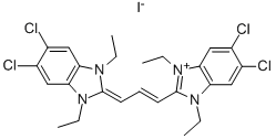
| CAS号 | 3520-43-2 |
| 分子式 | C25H27Cl4N4.I |
| 分子量 | 652.228 |
| EINECS号 | 200-258-5 |
| 熔点 | 275–278 ℃ |
| 溶解度 | 溶于甲醇、N,N-二甲基甲酰胺、二甲亚砜 |
| 形态 | 晶体 |
| 颜色 | 深红色 |
| 最大波长(λmax) | 514nm |
| 稳定性 | 自购买之日起,在 -20°C 下按供应状态干燥保存最多 2 年。 DMSO 或蒸馏水中的溶液可在 4°C 下储存长达 1 年。不要冻结/解冻。仅通过过滤对溶液进行灭菌,而不是通过高压灭菌 |
| 生物领域应用 | Detectingmitochondrialmembrane potential,ABCB1,ABCC1,and ABCG2 transporters inhibitors,nucleic acid hybridization,prostate cancer; treating cellular death,Alzheimer’s disease; apoptosis assay; cytotoxicity assay; hematotoxicity |
| 主要应用 | Langmuir-Blodgett films;lasing systems; nonlinear optical materials;photographic materials1, |
| WGK Germany | 3 |
Guidelines (Following is our recommended protocol. This protocol only provides a guideline, and should be modified according to your specific needs).
Labeling of Cells:
1. Culture cells in 6-, 12- , 24-, or 96-well plates at a density of 5× 10
5
cells/mL. Incubate the cells according to your normal protocol.
2. Ensure that the JC-1 and DMSO has equilibrated to room temperature, and then prepare a 200 μM stock solution by dissolving the contents of one vial in 230 μL of the DMSO provided.
3. For the control tube, allow the vial of CCCP has come to room temperature, add 1 μL of CCCP (50 mM). Incubate cells at 37°C for 5 minutes.
4. Add 10 μL JC-1 (200 μM) per well to make the final concentration at 2 μM. Incubate cells at 37°C, 5% CO
2
, for 15-20 minutes. If additional labeling followed, for example with an annexin V, begin with step 2.a. If not, proceed with step 1.e.
5. After incubation, centrifuge cells for 3-4 minutes at 400× g at 4°C, carefully aspirate the supernant.
6. Wash cells twice with PBS (1×): add 2 mL PBS (1×) to suspend cells and vortex to mix thoroughly. Centrifuge cells for 3-4 minutes at 400× g at 4°C, carefully aspirate the supernant.
7. Add 500 μL PBS (1×) to suspend cells. Analyze sample on a flow cytometer, fluorescence microscopy, or fluorescence microplate reader.
-
CAS号:7385-67-3
尼罗丝 -
CAS号:35935-34-3
碘化1-乙基-3-甲基咪唑 -
CAS号:150347-59-4
5(6)-羧基二乙酸荧光素琥珀酰亚胺酯(CFDA) -
CAS号:143-74-8
苯酚红 -
CAS号:3520-43-2
JC-1 -
CAS号:6478-79-1
5,6-二氯-2-甲基苯并咪唑 -
CAS号:3237-62-5
5,6-二氯-1-乙基-2-甲基苯并咪唑 -
CAS号:4333-62-4
1,3-二甲基咪唑碘盐 -
CAS号:7385-67-3
尼罗丝 -
CAS号:35935-34-3
碘化1-乙基-3-甲基咪唑 -
CAS号:150347-59-4
5(6)-羧基二乙酸荧光素琥珀酰亚胺酯(CFDA) -
CAS号:143-74-8
苯酚红 -
CAS号:3520-43-2
JC-1 -
CAS号:6478-79-1
5,6-二氯-2-甲基苯并咪唑 -
CAS号:3237-62-5
5,6-二氯-1-乙基-2-甲基苯并咪唑 -
CAS号:4333-62-4
1,3-二甲基咪唑碘盐